Catching up

So I felt like I couldn’t put off the sixteen hundred articles twiddling their thumbs and tapping their feet in my RSS reader any longer. This is the first part of the crop that has accumulated since late December (yikes!). Legless axolotls, homing starfish, secretly related proteins, and more!

1. Axolotls are good at regenerating – until you make them grow up.

(Portrait of a pale lab/aquarium variety axolotl by Orizatriz, Wiki Commons.)

It’s probably not exactly obvious from my posting record, but a large part of my PhD work is about regeneration. It’s something we humans are pretty shit at, but many other vertebrates aren’t. Axolotls, these adorably dumb-faced salamanders, can easily regrow their legs. However, lab axolotls are kind of permanent babies. Although they can grow up in the sense that they are able to breed, they normally keep larval characteristics like gills throughout their lives. It’s reasonable to suspect that this influences their regenerative ability – after all, tadpoles lose their ability to regrow limbs the moment they turn into frogs.

It’s possible to make axolotls metamorphose, too, if you treat them with thyroxine (the same hormone that induces metamorphosis in “normal” amphibians). And when they turn into proper adult salamanders, they suddenly become much poorer regenerators. They can still replace a limb – kind of. But they take twice as long as non-metamorphosed axolotls of the same age and size, and they invariably wind up with small, malformed limbs, often missing bones. After amputation, new skin is slower to grow over their wounds, and the cells that gather under the new skin are sluggish to divide. Something about metamorphosis – that isn’t simply age – dramatically changes how they respond to amputation.

Reference: Monaghan JR et al. (2014) Experimentally induced metamorphosis in axolotls reduces regeneration rate and fidelity. Regeneration advance online publication, doi: 10.1002/reg2.8

*

2. Similar cells repair muscles in crustaceans and vertebrates

“Regeneration” can cover a lot of different processes. For example, depending on the creature and the organ you’ve damaged, regenerated body parts can come from totally different kinds of cells. In planarian flatworms, a single kind of stem cell can replace anything else in the body. In the eyes of newts, mature cells of the iris transform into lens cells to replace a missing lens. In our muscles, there are special cells called satellite cells that are held in reserve specifically to make new muscle cells when needed.

This recent study of a little crustacean called Parhyale hawaiensis suggests that muscle regeneration in the fingernail-sized arthropod works in much the same way. Konstantinidis and Averof shot early embryos of Parhyale with DNA encoding a fluorescent marker, which randomly integrated into the genomes of some of the cells it hit. In a few “lucky” individuals, the marker ended up labelling just one cell lineage, and the pair used these animals to figure out which cells made which tissues in a regenerated limb.

It turned out that cells in Parhyale are limited in their potential. Descendants of the ectodermal lineage could make skin and nerves but not muscle, and the mesodermal lineage built muscle but not skin or nerves. Moreover, labelled cells only contributed to regeneration near their original location – animals with their left sides labelled never regrew glowing limbs on the right side. This is starting to sound a lot like vertebrates, but it’s still a very general observation. However, the similarities don’t end there.

Like vertebrate muscles, the muscles of the little crustaceans contain satellite-like cells derived from the mesodermal lineage that sit beside mature muscle cells and express the Pax3/7 gene. When the researchers transplanted some of these cells from animals with the glowy label into leg stumps of non-glowy animals, there were glowing muscle cells in some of the regenerated limbs. So like satellite cells, these cells can turn into muscle during regeneration. There’s little question that muscle cells have a common origin in vertebrates and arthropods like Parhyale, but it’s really cool to see that their mechanisms of regeneration also might.

Reference: Konstantinidis N & Averof M (2014) A common cellular basis for muscle regeneration in arthropods and vertebrates. Science, published online 02/01/2014, doi: 10.1126/science.1243529

*

3. Convergent evolution is a poor explanation of rhodopsins

Proteins can be difficult. I mean, sometimes they do their darnedest to hide their family ties. A protein is a chain of amino acids (on average about 300 of them) often folded into a complex shape. Closely related proteins have obviously similar amino acid sequences. However, more distant relatives can be harder to identify. There are about 20 different kinds of amino acids in proteins, so the number of possible sequences is unimaginably vast. The same function can be carried out by very different sequences, and therefore enough evolution can completely erase sequence similarity.

Protein structures are generally thought to be more conserved than sequences. Like function, structure allows for a huge amount of sequence variation without significantly changing. However, theoretically, it’s possible that two unrelated proteins have similar structures because of their similar functions, not because of common ancestry. Apparently, this has been argued for the two types of rhodopsins – proteins that harvest light in systems as different as a the “solar generator” of a salt-loving microbe and the photoreceptors of our own eyes.

If Type I and Type II rhodopsins are similar despite being unrelated, one would assume that this is because they need to be that way to capture light. There are, after all, astronomical numbers of possible protein structures, and the chances of two protein families accidentally stumbling onto the same one without selection steering are slim to say the least. But, in fact, you can rearrange the structure of a rhodopsin in all kinds of cunning ways without destroying its function. This rather weakens the case for convergent evolution, and suggests that similarity of structure does indicate common ancestry here.

Reference: Mackin KA et al. (2014) An empirical test of convergent evolution in rhodopsins. Molecular Biology and Evolution 31:85-95

*

4. Starfish can see their way back home

(Blue starfish, the beast featured in the paper, in its natural habitat. Richard Ling, Wiki Commons.)

Starfish aren’t widely known as visual creatures, but they do have eyes at the tips of their arms. The eyes are a bit… basic – no lenses, just a hundred or two little units filled with photoreceptors. Garm and Nilsson set out to find out how the starfish used their eyes. They measured or calculated the eyes’ visual fields (five arm-eyes together can see pretty much everywhere around the animal), resolution (very coarse), reaction speed (slow), and their sensitivity to various wavelengths (they are colour-blind, most sensitive to ocean blue).

Then they took some poor starfish and dumped them a little way off the coral reefs they like staying on. The creatures could walk home from short distances (about 2 m or less), but if you take them too far away, they just wander around in random directions. Likewise if you take off their eyes (don’t worry, they regenerate) or do the experiment in the dark. In conclusion: starfish eyes aren’t exactly top-end cameras, but they are definitely useful to the animals. And what would a slow, brainless mopper-up of coral reef rubbish do with eagle eyes anyway?

(The paper states the walking speed of these starfish as about 4-5 cm per minute. I have a feeling this wasn’t the most exciting fieldwork these guys have done…)

Reference: Garm A & Nilsson D-E (2014) Visual navigation in starfish: first evidence for the use of vision and eyes in starfish. Proceedings of the Royal Society B 281:20133011

*

5. What makes wormies settle

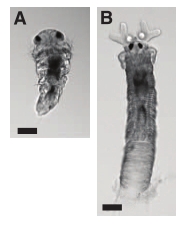
OK, Shikuma et al. (2014) one isn’t so much for its own news value, but I hadn’t known that my favourite worms need bacteria to settle until I saw this paper, so I think it deserves a mention. (Besides, it has beautiful pictures of baby Hydroides in it, which I couldn’t resist posting below. They are So. Cute. Yes, I’m weird.)

Shikuma_etal2014-hydroidesBabies

Tubeworms of the serpulid family have swimming larvae which are in many ways like the acorn worm larvae mentioned in my previous post (except cuter). They are tiny, look nothing like an adult worm, have bands of cilia for swimming and feeding, and live in the plankton until they’re ready to metamorphose. When they find a place they like, they settle and turn into adult worms. And apparently, this particular species (Hydroides elegans) not only needs a specific bacterium to like a place, it needs specific proteins produced by that bacterium.

The proteins in question are the components of a nasty device bacteria probably stole from viruses and then used to poke holes in one another. But to Hydroides larvae, they appear to be necessary for metamorphosis. Put healthy bacteria together with worm babies in a dish, and you’ll get happily settled little worms. Do the same with bacteria with damage to the relevant genes, and nothing happens. Use an extract containing the proteins but not the bacteria, and you still get metamorphosing worms. Use too much, though, and they start dying. Everything in moderation…

(Maybe my dismal failure at raising happy young worms years ago could have been remedied with the right bacteria?)

Reference: Shikuma NJ et al. (2014) Marine tubeworm metamorphosis induced by arrays of bacterial phage tail-like structures. Science 343:529-533

*

6. Relative of animals does strange multicellularity with familiar genetics

Although this idea probably hasn’t reached popular perception, animals are surrounded by other multicellular lineages in the tree of life. Sure, most of them are only part-time multicellular, but that’s beside the point. What’s clear is that multicellularity, at least in its simpler forms, is rampant in our extended family. Slime moulds do it, fungi do it, our closest relatives choanoflagellates do it, and our next closest relatives, filastereans and ichthyosporeans also do it.

These latter two groups are really poorly known (the fact that only a taxonomist could like the latter’s name probably doesn’t help), but the situation is getting better with the attention they are receiving as relatives of animals. There are now genome sequences out, and some people are looking at the life cycles of the little creatures to search for clues to our own origins.

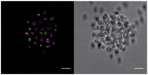
Iñaki Ruiz-Trillo recently published a paper describing an ichthyosporean that can form a weird kind of colony with many nuclei in the same membrane starting from a single cell (Suga and Ruiz-Trillo, 2013). Now his team describe a different kind of multicellularity in a filasterean, Capsaspora owczarzaki. Rather than developing from a single cell, this guy does something more akin to the slime mould way: take a load of individual cells and bring them together. (Below: a clump of Capsaspora cells from Sebé-Pedros et al. [2013]. On the right is a regular photograph of the colony. The two-coloured fluorescence on the left indicates that the colony formed by different cells coming together rather than a single cell dividing.)

Sebé-Pedros_etal2013-F4.capsasporaClump

But, interestingly, some of the genetics involved is similar to what animals use, despite the different ways in which the two groups achieve multicellularity. For example, we’ve known since all those genomes came out that the proteins animals use to glue cells together and make them talk to each other are often older than animals. Well, Ruiz-Trillo’s filasterean appears to ramp up the production of some of these when it goes multicellular. It also uses a gene regulation strategy that animals are really big on: it edits the RNA transcribed from many genes in different ways depending on cell type/life stage before it’s translated into protein.

A lot of the details are going to need further investigation, since this was a global RNA-sequencing study with a bird’s-eye view of what genes are doing. It’s still a nice reminder that, like most other innovations in evolutionary history, the multicellularity of animals didn’t spring fully formed out of nowhere.

References:

Suga H & Ruiz-Trillo I (2013) Development of ichthyosporeans sheds light on the origin of metazoan multicellularity. Development 377:284-292

Sebé-Pedros A et al. (2013) Regulated aggregative multicellularity in a close unicellular relative of metazoa. eLife 2:e01287

Animals, amoebae and plant scientists’ concerns

I recently wondered, in response to an “ideas” paper in BioEssays, whether animals, fungi, slime moulds etc. actually had a multicellular common ancestor. Dickinson and colleagues’ argument (partly) hinged on the shared presence of epithelia, “barrier” cell layers with distinct insides and outsides, in animals and the social amoeba Dictyostelium discoideum. The most recent crop from BioEssays includes a short letter by František Baluška of the botany department at the University of Bonn that challenges this argument.

Plants, Baluška reminds us, also have epithelia. These epithelia are functionally more similar to animals’ than the one Dickinson’s team found in the amoebae. While there may be doubts about amoebae, plants almost certainly became multicellular independently of animals. Ergo, convergent evolution can clearly produce similar tissues in two distant lineages. So why would we take the possession of an epithelium as evidence for a multicellular common ancestor?

Which is a perfectly valid argument, but it misses the point in my opinion.

The botanist writes,

[Plants] evolved their own plant-specific epithelia 3–5, obviously via convergent evolution. This fact alone not only continues to make plausible the traditional independent origin of multicellularity in the metazoa and social amoebae, but it also indicates that the power of convergent evolution should not be underestimated.

Of course it shouldn’t, but Dickinson’s team wasn’t arguing that “the traditional independent origin of multicelluarity” in animals and amoebae was not plausible any more. They find it unlikely that the functional and molecular similarity (does the latter exist between plants and animals?) between animal and amoeba epithelia is convergent, but they are suggesting that we investigate their new hypothesis, not that we summarily throw out the old one. Baluška is attacking a straw man.

Furthermore, he only addresses this one argument, but the thing in the Dickinson article that made me think the most was phylogeny. According to the traditional scenario, it seemed more likely that all those different unikont groups evolved multicellularity independently. But multicellularity is very widespread among unikonts, so precisely what makes the traditional scenario more likely? (Incidentally, has anyone done any actual statistics on this?)

As far as I’m concerned, the letter said nothing to change my mind. Dickinson et al. presented an interesting idea that’s definitely worth a closer look. I don’t think the evidence is currently strong enough to upset the consensus, but the proposal is not at all daft. I have to say I agree that plants should not be ignored, though. Because we can assume that any similarity between them and animals when it comes to being multicellular is the result of convergence, they’d be a wonderful “control group” when people start testing Dickinson et al.‘s hypothesis.

I think that’s something students of evolution should always keep in mind. Plants and animals have little reason to do things in the same way – they diverged very long ago, adapted to completely different lifestyles, etc. If they do so anyway, that might tell us something deeper about the way living things work. A limitation imposed by physics, a very ancient genetic predisposition, or simply the best way to do something – either way, finding the reason will enrich our knowledge of life and evolution. Animal scientists would be well advised to remember that.

***

Reference:

Baluška F (2012) Rethinking origins of multicellularity: Convergent evolution of epithelia in plants. BioEssays, available online 26/10/2012, doi: 10.1002/bies.201200134

Animals, amoebae and assumptions

Animals aren’t the only multicellular creatures in their phylogenetic neighbourhood. Social amoebae, many fungi and quite a few of the poorly known choanoflagellates spend at least part of their lives as collections of cooperating cells. Conventional wisdom has been that these groups invented multicellularity independently, but maybe conventional wisdom needs a bit of challenging.

To tell you the truth, I never really thought about the other possibility, that being multicellular is the original state of affair for these organisms. I never really considered the evidence on which the conventional wisdom was based. You could say I didn’t really care either way. A while back I saw a paper that said something about a social amoeba having an epithelium, but I just kind of shrugged and went on with my life. I don’t know, now an article in BioEssays brought this up again, and I’m not sure I was right to ignore it back then. I think Dickinson et al. (2012) have a point, and I think some assumptions may need to be reexamined.

In case you wondered, an epithelium is a type of tissue made of a layer or layers of polarised cells. “Polarised” means that various cellular components – proteins, attachments to neighbouring cells, organelles – are distributed unevenly in the cell, clustered towards one or the other side of the cell layer. Epithelia line pretty much everything in a typical animal’s body, from, well, the entire body, to things like guts and glands. They secrete important stuff like hormones, and their closely packed cells form a barrier to keep molecules and pathogens where they belong. An epithelium was thought to be a uniquely animal thing to have, but looking more closely at that weird little amoeba suggested it may not be.

The paper that I ignored was Dickinson et al. (2011) – yes, by the exact same people who wrote the BioEssays piece. OK, I didn’t completely ignore it. I read enough of it to scribble a quick note in my citation manager saying “screams convergent evolution to me”. The paper examined the multicellular stage in the life of Dictyostelium discoideum, an ordinarily single-celled amoeba that reacts to food shortages by crowding together with friends and family to form a fruiting body that helps disperse some of its cells in search of new habitats. The fruiting body is pretty complex for a “unicellular” creature, and it turns out that this complexity includes a region of tissue that looks quite a lot like a simple epithelium. It doesn’t just look like one; it sorts out its insides and outsides with the help of proteins called catenins, which are also involved in cell polarity in the epithelia of animals. (Below: D. discoideum being multicellular, from Wikipedia)

That isn’t much evidence to base an inference of homology on, especially since other key players in animal cell polarity are entirely absent from D. discoideum. But equally, the fact that tons of unikonts (the group including amoebae, slime moulds, fungi, choanoflagellates and animals) are single-celled doesn’t mean that the multicellular groups all came up with the idea independently. Evolution doesn’t always increase complexity – sometimes complexity becomes superfluous.

I remember when we discussed the choanoflagellate genome paper (King et al., 2008) in class. The genome in question belongs to a purportedly single-celled creature, but it contains tons of genes you’d think only multicellular organisms would need, such as genes for cell-to-cell adhesion proteins. So one explanation is that these proteins originally did something else, like anchoring a single cell to its favourite spot. Another explanation is that they did have something to do with multicellularity – it just wasn’t the multicellularity of animals at first.

This suggestion isn’t terribly controversial when you’re talking about choanoflagellates, since some of them do obviously form colonies (one such colony of Salpingoeca/Proterospongia rosetta is shown below, from Mark Dayel of the King lab via ChoanoWiki). It’s not hard to imagine that either the “single-celled” species whose genome was sequenced also has a colonial stage the scientists just never saw, or that its recent ancestors did.

Whether or not the same applies to the whole of unikonts is a more difficult question. I’m not at all familiar with the details of unikont relationships, but based on the tree shown in the BioEssays article, multicellularity is all over the group. In most cases, it’s facultative multicellularity; animals are rather the exception in being doomed to it for their entire lives. However, if you just looked at that tree, you’d wonder why the hell anyone thought the common ancestor of these things wasn’t some kind of multicellular.

Yet the details of animal-like multicellularity aren’t so widespread. True cadherins (the cell adhesion proteins I mentioned) have only been found in animals proper. Choanoflagellates and some even more obscure relatives of animals have bits and pieces of them, and other unikonts have none at all as far as anyone knows. Epithelium-like tissues have only been described in that one species of amoeba – but, as Dickinson and colleagues note, no one really looked in the others.

Personally, I wouldn’t be at all surprised if the conventional wisdom ended up shifting. I still don’t think that the evidence from Dictyostelium is enough to draw a conclusion. We obviously need to know a lot more about unikont genomes, tissues and life cycles to piece together the history of multicellularity in the group, but I’m not sure that right now a unicellular ancestor has a lot more going in its favour than a multicellular one. Guess we’ll have to wait and look with an open mind 🙂

***

References

Dickinson DJ et al. (2011) A polarized epithelium organized by β- and α-catenin predates cadherin and metazoan origins. Science 331:1336-1339

Dickinson DJ et al. (2012) An epithelial tissue in Dictyostelium challenges the traditional origin of metazoan multicellularity. BioEssays advance online publication, 29/08/2012, doi:10.1002/bies.201100187

King N et al. (2008) The genome of the choanoflagellate Monosiga brevicollis and the origin of metazoans. Nature 451:783-788


Bacteria invented multicellularity – then thought better of it

I get content alerts from a whole host of journals, some specialist publications focusing on my field, some more general. The majority of even the former is stuff I couldn’t care less about, and the ratio of interesting to irrelevant from generalist journals like PNAS or Nature is lower still. Nevertheless, sometimes you stumble on a title that isn’t directly related to your main interests, but still makes the whole rummaging through the Pile of Irrelevance worth it.

This was the case with a paper (Schirrmeister et al., 2011) just published in the online, open-access journal BMC Evolutionary Biology. It’s so fresh that they haven’t even formatted it – the full text is only available as a “provisional” pdf where all the figures are dumped at the back of the file, separated from their captions (why they can’t wait with publication until the damned thing is in readable format escapes me).

The study by Schirrmeister and others deals with an unusual group of bacteria. Cyanobacteria are probably still better known as blue-green algae, even though they have nothing to do with anything else we call an alga (well, in truth they have everything to do with algae, but in a rather more interesting way, as we’ll see below). If I had to pick one group of organisms that had the greatest impact on the history of our planet, cyanobacteria would be it. For more than two billion years, they have contributed huge amounts of biomass to the global carbon cycle. They are solely responsible for the oxygen-rich atmosphere of the earth – and, by extension, for most eukaryotic life and all animals. They are among the distinguished group of bacteria that can fix nitrogen – a vital ingredient of DNA and proteins – straight from the air, making it available for other organisms. Without them, the world would be a vastly different place, and we wouldn’t be possible. As Andrew Knoll puts it in his wonderful book Life on a Young Planet (consider this a recommendation ;)): “animals may be evolution’s icing, but bacteria are the cake”.

Cyanobacteria live everywhere there is light, from hot springs to the ocean to puddles to stone walls (as components of lichens). They also live inside the cells of every single eukaryote capable of photosynthesis: plants, red and green algae, brown algae, diatoms, dinoflagellates, euglenids (and any others I forgot to mention). The chloroplast is a pared down cyanobacterium – a symbiont that has lost most of its genes, but the ones that remain, together with its structure, still tell of its ancestry. Plants owe all their green splendour to these tiny buggers.

Cyanobacteria are not just immensely important, they are also quite unusual among prokaryotes. As the post title implies, they invented multicellularity. Multicellular cyanobacteria display a range of complexity. Some of them are just chains of identical cells. Others, though, have up to three different cell types. Heterocysts, thick-walled cells that ensure the oxygen-free environment that these bacteria require for nitrogen fixation, sit at regular intervals among “normal” cells, and when necessary, the “normal” cells can also differentiate into hardy resting cells that can survive bad times. The most complex cyanobacteria not only have filaments with different cell types, but also introduce branching into these filaments. This is the most complex prokaryotes get.

Filaments of an unbranched, differentiated cyanobacterium. The oversized heterocysts are quite obvious in some of them. Image by Kristian Peters, from Wikimedia Commons.

The new study raises an interesting possibility: that at least the simple form of multicellularity (i.e. undifferentiated filaments) occurred very early in the history of cyanobacteria. According to Schirrmeister et al., the vast majority of modern cyanobacteria descend from multicellular ancestors, even though a great many of them are single-celled today. Even more intriguingly, they find a lineage that might have re-evolved multicellularity after losing it. I don’t pretend to fully understand the methods used to come to these conclusions, but I have to say that it’s built on an impressive dataset – the group selected 58 cyanobacterial species for more detailed study from an original phylogenetic tree built from over a thousand taxa. They then constructed trees of this smaller dataset using two separate methods, and finally, tried to reconstruct the ancestral states at various points in those trees using several different statistical methods again. The analyses all agree: multicellularity is a very ancient trait in cyanobacteria, and it was lost left and right during their three-billion-year history.

These findings go against our ingrained view of evolution as an inexorable march towards increasing complexity. We, mammals, are among the (if not the) most complex organisms the earth has ever produced. We are assemblages of some 200 distinct cell types organised into a finely regulated machinery of a multitude of specialised organs. When we look at the large-scale patterns in the fossil record, we also see that this complexity has accumulated from much simpler beginnings over the aeons. We can be forgiven for thinking, in a characteristically self-centred way, that complexity is where evolution is intrinsically headed. But every now and then, nature reminds us that “more complicated” does not necessarily equal “favoured”.

Parasites are probably best known for their tendency to become simplified – after all, if you are bathed in your host’s digestion products all the time, why waste your energy on growing your own gut? However, simplification is abundant in organisms that make their own living, too. For example, two entire phyla of distinctly unsegmented, baglike worms – spoon worms and peanut worms -, likely came from more sophisticated segmented worms (Struck et al., 2007). Now, cyanobacteria join the club, and new questions surge in their wake. Why did they go back to unicellularity? How difficult is it for them to become multicellular? Such questions, of course, can be asked about any complex trait that followed a similar evolutionary trajectory.

Most intriguingly, these tiny microbes seem to violate another “law” of evolution, known as Dollo’s law: that once lost in a lineage, a complex trait won’t reappear. If the inferences of Schirrmeister et al. are correct, then either simple multicellularity isn’t such a big deal at all for these bacteria, or Dollo’s law isn’t as much of a law as we thought.

(Actually, the latter is probably the case however the history of cyanobacteria turns out. Dollo’s law has been questioned by others, and it was recently dealt a spectacular blow by a frog that almost certainly re-evolved teeth in its lower jaw (Wiens, 2011) after at least 200 million years of not having them.)

Evolution is a fascinating story. As the example of cyanobacterial multicellularity suggests, it can also be as complex as any good novel. I for one think this makes for a much more interesting and fulfilling narrative than simplistic listings of “what’s new” through the ages.

– – –

References:

Schirrmeister BE, Antonelli A and Bagheri HC (2011) The origin of multicellularity in cyanobacteria. BMC Evol Biol 11:45

Struck TH et al. (2007) Annelid phylogeny and the status of Sipuncula and Echiura. BMC Evol Biol 7:57

Wiens JJ (2011) Re-evolution of lost mandibular teeth in frogs after more than 200 million years, and re-evaluation of Dollo’s Law. Evolution advance online publication, DOI: 10.1111/j.1558-5646.2011.01221.x