Random of the day

Because productivity is too much effort. In my defence, it was paper writing-related curiosity that led me to Wikipedia, where I found this electron microscope image of a broken piece of mother of pearl/nacre by Fabian Heinemann. (In case you wondered, I wanted to check roughly how big nacre tablets were. And no, Wikipedia is not my only source for this ;)) So: this is what mother of pearl looks like when you zoom in a few thousand times.

Nacre is made of little tablets of aragonite stacked on top of one another and separated by sheets of organic matter. The way the tablets scatter light is what gives pearls their pretty, pretty shine.

(I have a thing for electron micrographs of biominerals. Actually, I’m a big fan of close-up images of pretty much anything. It’s like looking into the secret heart of things.)

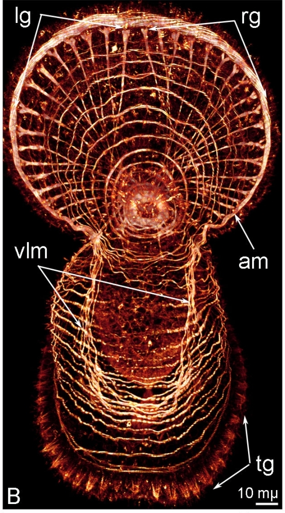
Postcard from crazyland

Contrary to all appearances, this is completely real. O.O

Temereva_Tsitrin2013_3b

What you are looking at is the fluorescently stained muscles of a phoronid larva. (Phoronids are one of the zillion variations on “tentacled filter feeder sitting in a tube” nature has come up with. They are related to lamp shells.) I couldn’t care less about larval muscles – not even sure why I opened the paper – but that looks friggin’ awesome. And a bit psychedelic.

(Source: Temereva & Tsitrin (2013), BMC Developmental Biology 13:14)

How cool is THAT?

I went to a talk by Paul Brakefield today. I loved it. It was a summary of decades of work on butterfly eyespots covering just about everything about them. There was development and evolvability, ecology and adaptation, courtship and speciation, really all an evolutionary biologist could wish for. Yay!

Nonetheless, my biggest squee moment in the entire talk was the first slide, which contained a picture of Kjell Sandved’s butterfly alphabet.

Back in the sixties, this guy who had taken like one photo in his entire life realised he could spell things out with patterns on butterflies’ wings. So he went and learned photography and travelled the world to hunt down the entire Latin alphabet plus symbols and Arabic numerals. All on little scaly lepidopteran wings. I have no words for how fucking cool that is. I don’t want to post the alphabet itself since it’s copyrighted, but do a google search or go here and you’ll see. You’ll see.

Wow.

Velvet worms in a prettier light

I bumped into Mayer et al. (2010) while hunting for reagents to use in an experiment I’m planning. The article is about segmentation (sort of), so I had to have a closer look, and man. Those pictures. Fluorescence and a good microscope can do wonders. This is what a velvet worm looks like in normal light (whole body shot of an unspecified peripatid by Geoff Gallice, Wikipedia, and portrait of Euperipatoides rowelli by András Keszei via EOL):

I think they are adorable and cuddly the way they are (apart from that hunting with slime bit), but they look simply gorgeous if you stick some glowing antibodies to them and start playing with a confocal ‘scope.

This is a fairly late-stage embryo of E. rowelli, the same guy waving its chubby leggies at you in the right-hand photo above. The green dots are cells that were copying their DNA when the baby was killed (all of the pictures below are from Mayer et al., of course):

These are younger embryos of the same species, with all their DNA labelled in blue and dividing cells labelled in red:

And these are embryos of another species from the same family as the unidentified guy from Wikipedia (colours are the same as above):

Seriously, there is something about the mystical glow of these images that always gets me. I think you could make almost anything look beautiful with a fluorescent marker and the right equipment. I know aesthetic appeal isn’t the primary aim of scientific imaging, but damn. Look at those alien creatures glowing with the light of the unknown.

In case you wondered, the point of the paper is that velvet worms lack a posterior growth zone. That means that when they develop their numerous segments, there isn’t a well-defined pool of cells at the rear of the embryo that divide to generate segment material. As you can see in all the red glow, cell division happens evenly all over the place. Why is this significant? Well, posterior growth zones were thought to be one of the characteristics that segmented animals might have inherited from their common ancestor. But Mayer and colleagues point out that the existence of a PGZ in the arthropod ancestor is dubious at best, and velvet worms (one of the closest living relatives of arthropods) also lack one, so maybe it’s kind of wrong to use the PGZ as an argument for the common ancestry of segmentation.

(There, that’s the science in a nutshell. Now I’ll just go back and admire the pretty glowy pictures some more :D)
***

Reference:

Mayer G et al. (2010) Growth patterns in Onychophora (velvet worms): lack of a localised posterior proliferation zone. BMC Evolutionary Biology 10:339

Goin’ a-hunting

It’s a little-known fact that before/in between wanting to be a biologist, I almost got sucked into astronomy. The cosmos still fascinates me, from the menagerie of space rocks and gas balls that fill our own solar system to the mysteries at the edge of the known universe. To the evolutionist in me, the possibility of life on other worlds is an especially tantalising idea. And now we are finding other worlds at a breakneck pace. I don’t think we will ever know what life is like on any of them, though detecting its existence may once become possible.

Did I mention planet hunting is awesome?

I am talking about the citizen science project Planet Hunters, of course. This is only one of the amazing projects you can participate in at the Zooniverse (which gets its name from Galaxy Zoo, the project that started it all). The main mission of Planet Hunters is, of course, to find planets orbiting other stars. You, the user have to look at a month’s worth of brightness measurements from a star, and search for the tell-tale dips that betray an extra-solar eclipse. Like this:

Most of the more spectacular ones have already been found by this point – either by your fellow hunters, or by the team operating the Kepler space telescope, which provides all the data. However, there are so many other gems to discover among those messy light curves that it almost doesn’t matter if your planet-hunting thunder is perpetually stolen.

Sometimes, you find pure beauty. One of the most common types of Interesting Stuff that the Kepler data offer is eclipsing binaries. These are pairs of stars orbiting each other in a way that we see their orbits edge on. Like the planets, these binaries eclipse their companion stars. Since stars are bigger and brighter than planets, the eclipses are much bigger compared to the noise in the data, so an EB has neat, clean dips in its light curve, occurring with clockwork regularity.

Some of them are so close together and orbit so fast that at Kepler’s resolution, a month of their light looks more like lace than a pattern of ups and downs.

And then there are all the others; dwarfs and giants, variable stars regular and haphazard, huge flares, weird things like cataclysmic variables. Even if you are in it for the planets, you can’t help but learn a lot about the stars. After a while, they become like family. You look at a light curve and you can immediately guess whether it’s a dwarf or a giant, whether it’s cool or hot, whether it’s a binary or a loner, or even if its’s one of the rarer breeds of stars you might come across. It’s a bit like birdwatching. If you’ve ever got disproportionately excited from recognising a rare bird (or flower, or insect, or sports car), you know what I mean. (If you haven’t, what are you waiting for? ;))

I’m grateful to the people who make these adventures possible. It’s great that I can play at astronomy, see all that neat stuff, contribute to a field I have absolutely no expertise in, and learn from the knowledgeable folks that hang around the forums. The Zooniverse deserves every one of its hundreds of thousands of users and millions of clicks, is all I’m saying 🙂
Pretty Reapers

Did you know proteins were beautiful?

Of course, many of them are just vaguely potato-shaped lumps of joined atoms. But when a few of these lumps get together, the most amazing shapes can emerge. Such as these:

Structural models of different forms in various views

What the molecule actually looks like

These are models of a fruit fly apoptosome, a huge multi-protein complex that plays a role in programmed cell death. (Programmed cell death, or apoptosis, is not as bad as it sounds. It’s used to remove damaged cells, to sculpt body parts during development, and other decidedly useful things.) The top image shows the structure of the apoptosome in the stylised language of molecular biology – spirals, ribbons, loops etc. indicate the way the protein is folded in that region. The bottom image gets closer to what the molecule would look like, although what the blue shapes represent – electron clouds – are kind of hard to picture in a realistic way. (The pictures are from Yuan et al. (2011) Structure of the Drosophila Apoptosome at 6.9 Å Resolution. Structure 19:128-140)